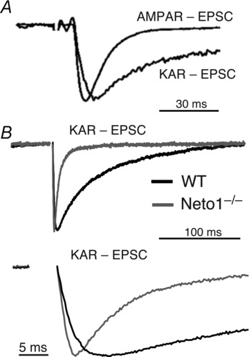
Figure 2. Distinct kinetics of native KARs in the brain.

A, in CA3 pyramidal cells of the hippocampus, mossy fibre activation elicits a fast kinetics AMPAR-EPSC and a distinct slow kinetics of KAR-EPSC. These responses have been scaled to the peak of the AMPAR-EPSC. The KAR-EPSC was recorded in the presence of the AMPAR selective antagonist GYKI53655. From Castillo et al. (1997); reprinted by permission from Macmillan Publishers Ltd: Nature©1997. B, in Neto1 knockout mice, both rise time and decay kinetics of mf-CA3 KAR-EPSCs are substantially accelerated. Normalized KAR-EPSCs from wild-type and Neto1 knockout mice are superimposed and depicted at a slow (top panel) and fast (bottom panel) time base. From Straub et al. (2011a); reprinted by permission from Macmillan Publishers Ltd: Nature Neuroscience©2011.
