
Figure 3. Distinct distribution of KARs in the brain.
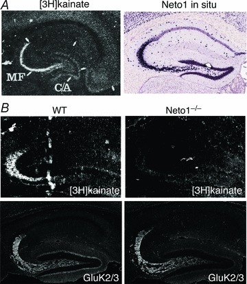
A, [3H]kainate signal shows distinct distribution of high-affinity kainate receptors in the hippocampal stratum lucidum, where mossy fibre (MF) and CA3 pyramidal cells form synapses Modified from Monaghan & Cotman (1982); by permission from Elsevier: Brain Research©1982. Neto1 mRNA is strongly expressed in CA3 pyramidal cells. Image from Allen Brain Atlas. B, [3H]kainate signal was reduced in Neto1 knockout mice (Neto1−/−), whereas GluK2/3 localized at synapses, suggesting that Neto1 only modulates [3H]kainate affinity, but not KAR synaptic localization. Modified from Straub et al. (2011a); reprinted by permission from Macmillan Publishers Ltd: Nature Neuroscience©2011.