
Figure 1. Whole-head mouse slices preserve peripheral and central brainstem auditory stations.
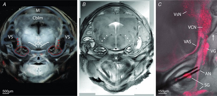
Selective expression of fluorescent reporter protein in an embryonic mouse whole-head slice preparation highlights auditory and vestibular system structures. A, coronal tissue slice, using transmitted light, of entire mouse head at E17-containing cochleae (dashed red outlines), auditory nerves (red arrowheads), peripheral vestibular system (VS), brainstem region (BS), superior olivary complex (SOC), midbrain (M), cerebellum (Cblm) and oral cavity region (OC). B, same tissue from panel A visualized with DIC optics to better reveal brain fibre tracts; arrowheads identify 7th cranial nerve roots. C, confocal image stack projection shows distribution of td-Tomato expression under control of parvalbumin promoter, overlayed on same tissue slice imaged with DIC optics. At embryonic ages, fluorescence is restricted to spiral ganglion (SG), auditory nerve (AN), vestibular ganglion (VG), ventral cochlear nucleus (VCN) neurons, vestibular nuclei (VsN) and axons of the ventral acoustric stria (VAS). Bipolar stimulating electrodes (black) are positioned on the AN, and recording electrode (arrow) is positioned in the ventral region of the VCN near auditory nerve entry zone.