
Figure 5. Electrophysiological properties of developing VCN neurons.
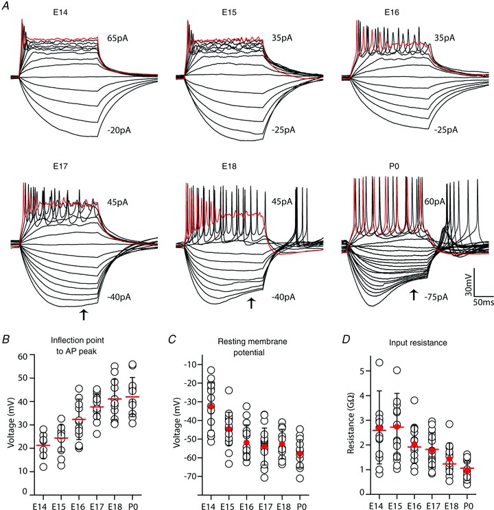
A, representative voltage responses of VCN neurons to serial injection of current steps (5 pA increments, 200 ms duration). Range of plotted current step values are reported for each series; some traces at larger positive values removed to clarify presentation. Voltage response to largest plotted depolarizing current step shown in red. Small AP responses followed by a steady depolarization are present in some cells at E14 and E15. Later ages show progressive appearance of tonic AP firing patterns during current step and sag (arrows) in hyperpolarizing voltage. B, AP amplitude, measured as difference between peak and inflection point, increased with VCN neuron maturation from 21 mV at E14 (n = 7) to over 40 mV by P0 (n = 11–14, each remaining age). C, resting membrane potential (RMP) for VCN neurons declines from –32 mV to about –60 mV by P0 (n = 15–19, all ages). D, input resistance at RMP (filled red circles) declines from very large values (average 2.7 GΩ) after E15 (n = 15–19, all ages). Red bars indicate average values with cells at standardized membrane potential (SMP, –73 mV), open black circles indicate values for individual neurons and black error bars show SD.