
Figure 6. Spiral ganglion neurons generate action potentials by E14.
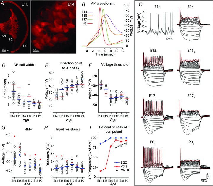
A, wide field view of an E18 cochlea (left) in Pv-tdTomato mice shows fluorescent inner and outer hair cells (HC), spiral ganglion (SG) and auditory nerve (AN) fibres. By E14, SG cell bodies and proximal neurites can be observed with fluorescence imaging (right), facilitating their identification within the cochlea for electrophysiological recording. B, representative AP waveforms of developing SGCs evoked by depolarizing current injection at SMP. C, spontaneous APs occur in SGCs at E14 (upper left) and older ages (not shown). Voltage responses to serial injection of current steps (5 pA increments) are illustrated by representative cases at E14, E15, E17 and P0. AP response is phasic at E14, but can be tonic (subscript 1) or phasic (subscript 2) in older SG cells. Hyperpolarizing current can convert tonic to phasic AP pattern (E15 subscripts 1 and 2 are the same neuron). Red trace is response to largest depolarizing current. D–F, APs become faster and larger, and voltage threshold becomes hyperpolarized during this developmental time frame. G, resting membrane potential becomes more hyperpolarized with age. H, input resistance at RMP (filled blue circles) does not change dramatically across this age range and is similar for SMP (blue bars). In panels D–G, average values for VCN neurons are depicted for comparison (red bars). I, as a population, SGCs acquire AP capability prior to VCN neurons, and VCN neurons prior to MNTB neurons (latter data re-plotted from Hoffpauir et al. 2010, asterisk). In panels D–H, open black circles indicate values for individual neurons, blue denotes SGC values, red denotes VCN values, bars indicate averaged SMP values, filled circles indicate averaged RMP values and black error bars show SD.