
Figure 11. dINs and the start of swimming.
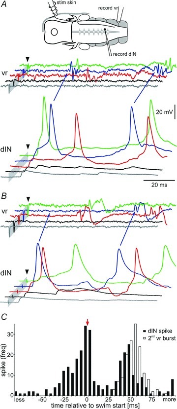
A and B, two examples of dINs receiving excitation after head skin stimulation (arrowhead) at increasing strength (from bottom to top trace grey via black, red and blue to green). EPSPs in dINs sum both spatially and temporally and can lead to spike firing and the start of swimming seen in the ventral root (vr, arrows blue trace). B, in one case (arrows in blue trace) the first dIN spike is NOT followed by a vr burst, but the second spike is. Grey bars mark the stimulus artefacts (cropped). C, histogram showing dIN spike timing (black bars) relative to the first ventral root spike, which marks the start of swimming (t = 0; arrow). The open bars show the time to the start of the second vr burst (second cycle of swimming). dIN firing is variable but generally occurs just before ventral root activity. Data are from 150 swimming episodes from 12 dIN recordings, bin size: 5 ms.