
Figure 5. The spontaneous synaptic activity recorded in rd1 ganglion cells in the presence of blockers of Ih, Na+ channels or gap junctions.
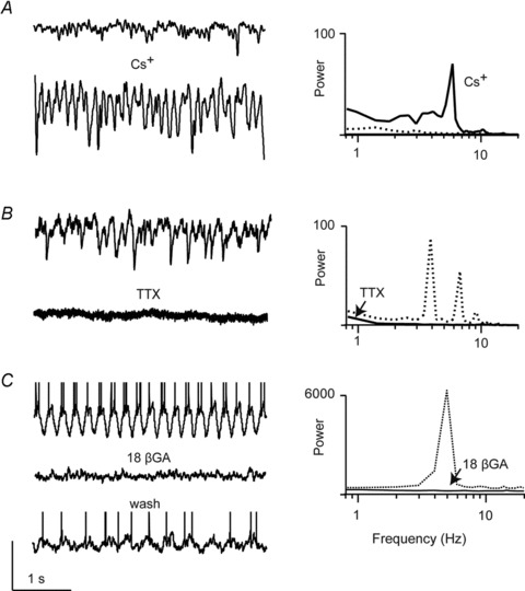
A, sEPSCs in rd1 ON ganglion cells recorded in control Ringer solution (Vhold∼−60 mV; top panels) and in the presence of 1 mm Cs+ (bottom panel) or B, 1 μm TTX (bottom panel, recorded in a different ganglion cell). C, oscillatory activity recorded in current-clamp mode (spikes are truncated to emphasize baseline membrane oscillations) from a ganglion cell in control Ringer solution (top panel) or in the presence 25 μm of the gap junction blocker 18β-glycyrrhetinic acid (18β-GA; middle panel). Bottom panel shows wash. The power spectra of these traces are shown on the right (activity measured in control Ringer solution is shown as dotted traces; those measured in drugs are shown as continuous traces). Vertical scale bar: 100 pA (for A and B), 40 mV for C.