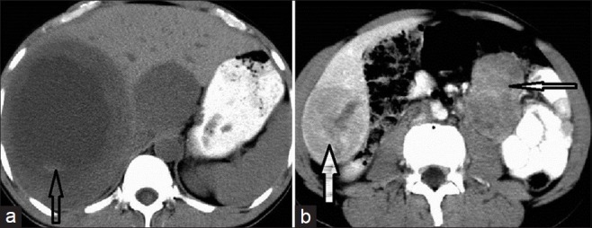
Figure 1.

NECT of a small bowel GIST with liver metastasis in a 42-year-old male. (a) Axial scan shows two well-defined, hypodense, space-occupying lesions in the liver. There is central necrosis. A small hyperdense focus within it is suggestive of a bleed (open arrow). (b) The primary mass shows heterogeneous enhancement (thin white arrow); another heterogeneously enhancing SOL can be seen in segment VI of the liver (solid thick arrow).
