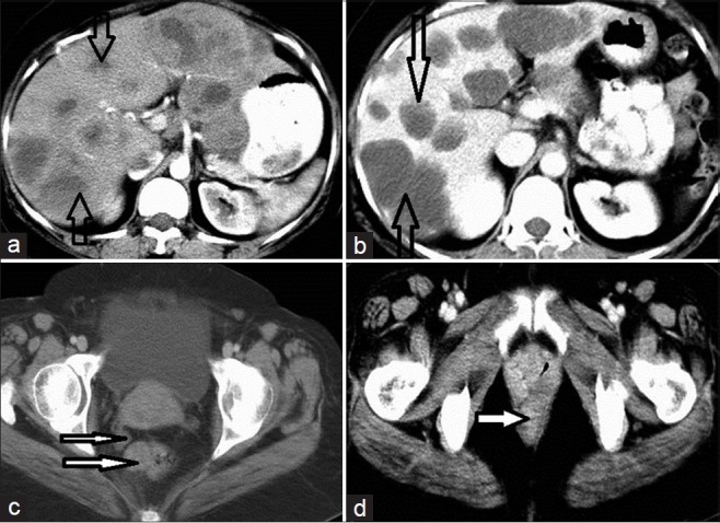
Figure 3.

CECT of the abdomen in a 45-year-old female patient shows rectal GIST with metastasis, before and after treatment. (a) Scan before treatment shows multiple solid enhancing SOL of varying sizes scattered in both lobes of the liver (open arrows). (b) After treatment the lesions are more hypodense, similar to the cyst, which is characteristic of metastatic deposits (two solid arrows). (c) The thick white solid arrow indicates rectal GIST; the lesion appears as a solid enhancing exophytic growth from the rectum; the thinner arrow indicates deposit in the pouch of Douglas. (d) After treatment the rectal GIST disappears and no mass is visible at the primary site (arrow).
