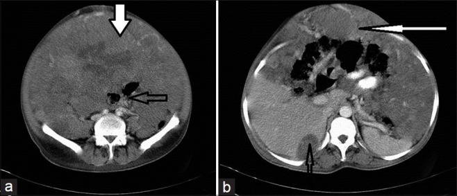
Figure 5.

CECT of abdomen shows extensive peritoneal dissemination in a 45-year-old male. (a) Scan shows the entire peritoneal cavity is filled with solid enhancing SOL (thick solid arrow), displacing and compressing the mesentery and bowel loops (open arrow). (b) Scan shows scalloping of the liver, caused by a peritoneal deposit (thin open arrow); Also shows omental caking (thin white arrow) that mimics pseudomyxoma peritonei.
