Figure 4.

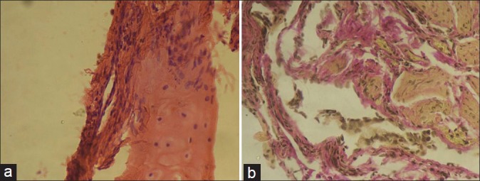
(a) Transbronchial lung biopsy (hematoxylin and eosin stain) showing fibroblasts adjacent to the bronchial cartilage (arrow). (b) Transbronchial lung biopsy (Van Gieson's stain) showing multiple areas of collagenous tissue (stained in pink color) and fibroblasts (elongated cells with brown-colored nuclei).
