Figure 1.

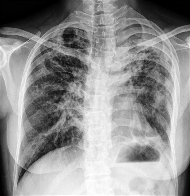
Plain chest radiograph (PA view) showing extensive bilateral fibrosis, more on the left side with a right upper zone cavity. A few cystic shadows were seen in both lungs

Plain chest radiograph (PA view) showing extensive bilateral fibrosis, more on the left side with a right upper zone cavity. A few cystic shadows were seen in both lungs