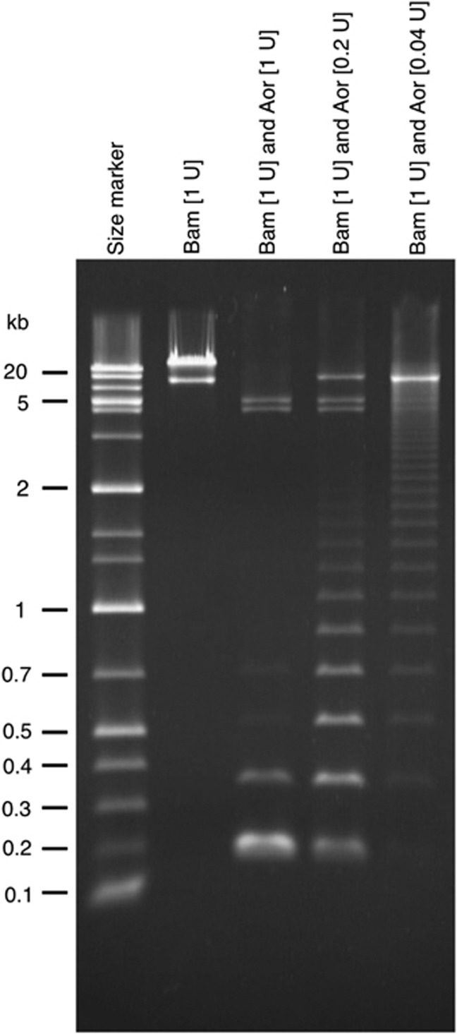
Figure 3.
Repetitive sequence structure revealed by restriction enzyme digestion. The second lane from the left (complete digestion with BamHI) shows two DNA fragments, the lower (8.1 kb) and upper (∼40 kb) bands being the vector (pCC1FOS) and insert (genomic DNA fragment of siamang), respectively. The third lane (complete digestion with BamHI and Aor51HI) contains two bands from the vector (split into two fragments of 4.2 and 3.9 kb due to an internal Aor51HI site) and other small fragments originating from the insert DNA. The prominent band at about the 0.2-kb position, which is absent in the second lane, indicates that the insert DNA digested with the two enzymes consists of a large number of restriction fragments (generated by Aor51HI digestion) of this size. The other two lanes contain the products of partial digestion with Aor51HI, and the appearance of ladder patterns indicates the presence of tandemly repeated sequences in the insert DNA.

