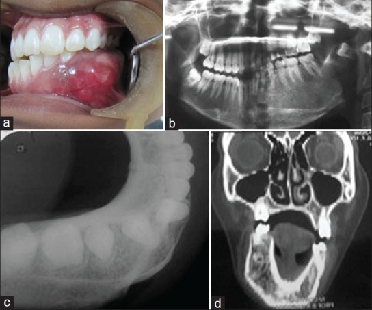
Figure 1.

(a) Preoperative intraoral view of the lesion; (b) Panoramic radiograph showing a radiolucent lesion extending from left ramus to right parasymphyseal area with impacted a third molar; (c) Occlusal radiograph showing the expansion of the buccal and lingual cortical plate; (d) Computed tomographic image showing expansile lesion
