Figure 2.
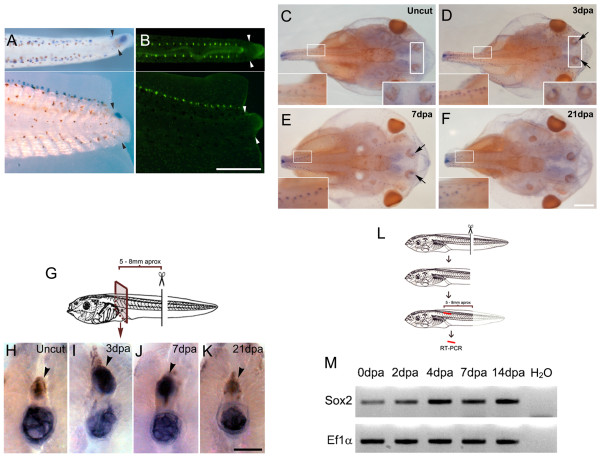
Systemic upregulation of Sox2 after tail amputation. (A,B) Tails from stage 49 tadpoles at 3 days post amputation (dpa). Dorsal (top) and lateral (bottom) view. (A) sox2 in situ hybridization and (B) 2-(4-(dimethylamino)styryl)-N-ethylpyridinium iodide (DASPEI) vital dye staining to identify the lateral line. Arrowheads indicate the amputation plane. (C-F) Dorsal view of sox2 whole mount in situ hybridization of the cephalic region of (C) non-amputated tadpoles, and tadpoles at (D) 3, (E) 7 and (F) 21 dpa. Insets show higher magnification of the lateral line and olfactory epithelial region that increase sox2 expression at 3 and 7 dpa. Notice that after fixation, tails were removed from their base and the observed stump ends do not correspond to the original amputation. (G) Diagram depicting the experimental set up for (H-K). (H-K)sox2 in situ hybridization in areas 5 to 8 mm rostral to the amputation plane from tadpoles at different dpa. sox2 is present in the spinal cord at 3 and 7 dpa (indicated by arrowheads). Endogenous expression of alkaline phosphatase in the notochord has been described previously [27] suggesting that expression on this tissue is non-specific. (L) Diagram depicting the experimental set up for (M). (M) Reverse transcription polymerase chain reaction (RT-PCR) analysis of sox2 mRNA levels in isolated spinal cord indicates that sox2 is upregulated in a rostral region during tail regeneration. EF1α was used as loading control. Scale bars: (A,B) 400 μm, (C-F) 400 μm, (H-K) 100 μm.

