Figure 1.
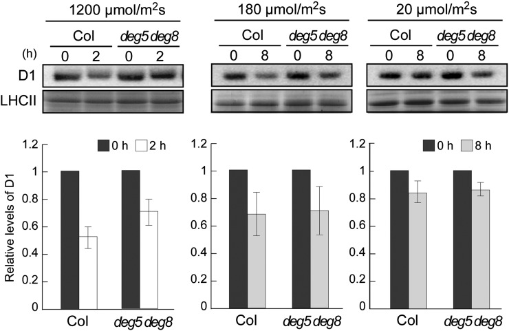
Immunoblot analysis of D1 protein in the deg5 deg8 mutant under three light conditions. Detached mature leaves of Col and deg5 deg8 (approximately 6-week-old plants grown under normal conditions) were preincubated with 5 mm lincomycin. The leaves were incubated for 2 h under high-light conditions (1,200 µmol photons m−2 s−1) or for 8 h under growth-light and low-light conditions (180 and 20 µmol photons m−2 s−1, respectively). Representative immunoblots (normalized by fresh weight) using anti-D1 (C-term) antibodies and the bands corresponding to Coomassie Brilliant Blue-stained LHCII are depicted. Signals of immunoblots were quantified using the ImageJ program and were normalized to the amount of Coomassie Brilliant Blue-stained LHCII (error bars indicate sd; n = 3). To compare D1 levels, ratios at 0 h were adjusted to 1.

