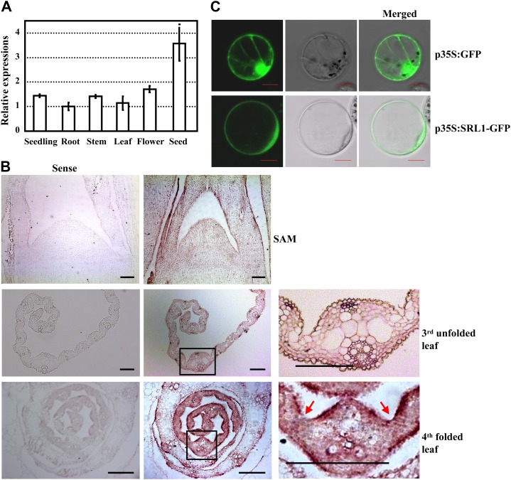
Figure 4.
Expression pattern of SRL1 and subcellular localization of SRL1. A, qRT-PCR analysis of SRL1 expression in various tissues including 14-d-old seedlings (whole plants without roots), roots, stems, 10th leaves at the heading stage, flowers, and seeds (9 d). Transcript levels were normalized with the ACTIN transcription and relative expression levels were compared with that in roots (set as 1). Mean values were obtained from three independent experiments and error bars indicate sd. B, In situ hybridization analysis of SRL1 expression in shoot apical meristem, the third unfolded leaves, and the fourth folded leaves of 14-d-old seedlings. SRL1 transcript was detected throughout the leaf primordium and unfolded leaves except that the signal was less intense in the bulliform cells. The SRL1 signal was more intense in the epidermal cell layers of folded leaves in the leaf sheath except for the region where sclerenchymatous cells are formed and the hollow region where bulliform cells are formed (red arrows). The squared regions (black boxes) in the middle section are enlarged to highlight the SRL1 expression (right section). Bars = 100 μm. C, Transient expression of SRL1-GFP fusion protein in rice protoplasts revealed that SRL1 is mainly located at the plasma membrane. Rice protoplasts expressing GFP alone were used as the control. Bars = 10 μm.

