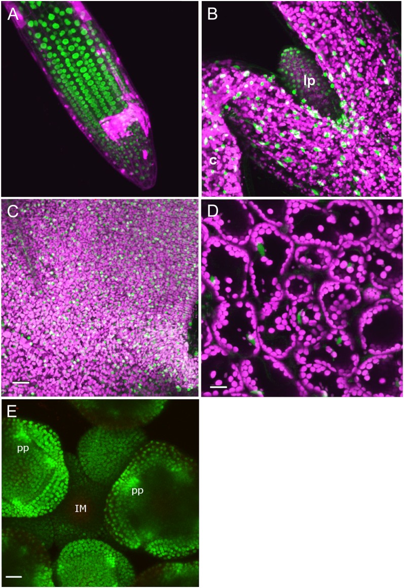
Figure 1.
Localization of TCP20-GFP in different plant tissues visualized by confocal laser scanning microscopy. TCP20-GFP signal is depicted in green, and autofluorescence of plastids is depicted in magenta. TCP20-GFP signal is mainly nucleus localized and can be detected in particular cell lineages of differentiating roots (A), the first leaf primordia in 3-d-old seedlings (B), the majority of leaf cells of the first initiated leaf, 14 d after germination (C), leaves 28 d after germination (D), and young flower buds at different developmental stages (E). Note that in the young floral meristems, strong expression is observed in the anlagen for petal primordia. c, Cotyledon; IM, inflorescence meristem; lp, leaf primordium; pp, petal primordium. Bars = 25 µm.

