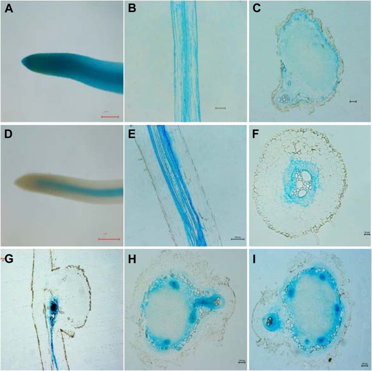
Figure 4.
Observation of GUS staining in transgenic soybean roots and nodules harboring the GmPT5 promoter::GUS fusion. A to C, Expression of empty vector with 35S promoter in roots (A and B) and nodules (C). d to F, Expression of proGmPT5::GUS in roots without rhizobium inoculation. D, Root tips. E and F, Longitudinal section (E) and cross-section (F) of mature roots. G to I, Expression of proGmPT5::GUS in nodules. G, Conjunction region between nodule initial and root at an early stage of nodulation. H and I, Intermediate (H) and mature (I) nodules. Soybean transgenic plants were grown in low-N and low-P nutrient solution for 15 d (G) and 30 d (A–F, H, and I). Bars = 500 μm for A and D, 20 μm for F and G, and 100 μm for all other images.

