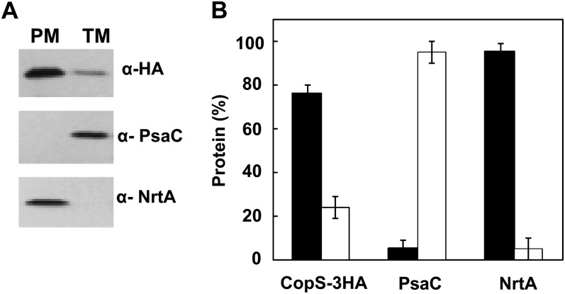
Figure 7.
CopS is localized to plasma and thylakoid membranes. A, Membrane localization of CopS. Membrane fractions from COPSHA strain induced for 4 h with 2 μm of nickel were prepared by Suc density gradient and aqueous polymer two-phase partitioning. Five micrograms of total protein were loaded and separated by SDS-PAGE. CopS-3HA, NrtA, and PsaC proteins were detected by western blot. PM, Plasma membrane; TM, thylakoid membrane. B, Quantification of CopS in different membrane fractions. Western-blot signal of three independent experiments were quantified using Image J program and averaged; error bars represent se. Plasma membrane (black); thylakoid membrane (white).

