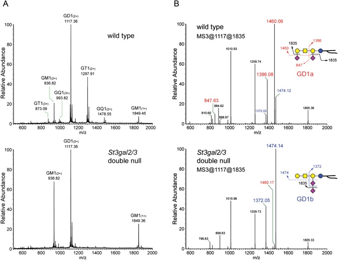
Fig. 4.
MS of permethylated wild-type and St3gal2/3-double-null mouse brain gangliosides. (A) Region of the full MS showing major brain gangliosides, with GT1 and GQ1 species diminished in St3gal2/3-double-null mouse brain gangliosides. (B) MS3 analyses of the permethylated disialoganglioside peaks of wild-type and mutant mouse brains demonstrating the relative intensity of GD1a-related fragments (red labels) and GD1b-related fragments (blue labels).

