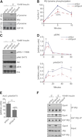
FIG. 3.
Gpc4 regulates insulin receptor activation and downstream signaling. A: Western blots from insulin- and IGF1R β−subunit immunoprecipitations of confluent shScr and shGpc4 preadipocytes, blotted for insulin/IGF1R β and pTyrosine before and after 5 min of 10 nmol/L insulin stimulation. B: Quantification of tyrosine phosphorylated insulin receptor in 3T3-L1 preadipocytes, normalized to total insulin receptor levels (n = 6). C: Western blots of confluent shScr and shGpc4 preadipocytes from total cell lysates before and after 5-min stimulation with 10 nmol/L insulin. D: Quantification of ERK and AktS473 phosphorylation at 0, 5, 10, 20, 40, and 60 min after insulin stimulation. pERK and pAktS473 were normalized to total ERK and Akt levels (n = 8). E: Area under the curve of AktS473 phosphorylation shown in D. F: Coimmunoprecipitation of Gpc4 with insulin and IGF1R β-subunit in 3T3-L1 cells. For all stimulation experiments, confluent undifferentiated preadipocytes were serum-starved for 3 h and stimulated with 10 nmol/L insulin. **P < 0.01; ***P < 0.001. (A high-quality color representation of this figure is available in the online issue.)

