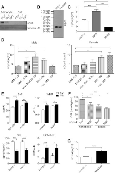
FIG. 5.
Gpc4 is released from adipocytes and correlates with markers of body fat and insulin resistance. A: Western blot for Gpc4 from conditioned serum-free Opti-MEMI of cultured isolated subcutaneous, perigonadal, and brown adipocytes and the corresponding SVF. Ponceau-S staining shows equal loading of proteins. Cells were isolated by collagenase digest and medium was conditioned for 12 h. B: Western blot of serum Gpc4. Glycoproteins from serum of 4-month-old C57BL/6 male and female mice were purified using anion exchange chromatography. Western blots from concentrated eluates were probed for Gpc4. C: Gpc4 ELISA from serum of C57BL/6 mice fed an HFD for 8 weeks, ob/ob and control mice. Control mice are C57BL/6 chow diet–fed mice and ob/+ mice combined (n = 6 per genotype). D: Gpc4 ELISA from serum of nondiabetic females (n = 77) and males (n = 83) grouped according to BMI and body fat distribution. Visceral overweight and obesity is defined by a CT or MRI ratio >0.4 between subcutaneous and visceral fat areas. E: Comparison of BMI, WHR, and GIR during a euglycemic hyperinsulinemic clamp and HOMA-IR of the lowest and highest quartile of serum Gpc4 levels of females and males (n = 19 and 20 per quartile, respectively). F: Comparison of GIR from nonobese (BMI <30) and obese (BMI >30) subjects divided into groups with low serum Gpc4 levels (≤5 ng/mL) and high serum Gpc4 levels (≥9 ng/mL). G: Serum Gpc4 levels in 30 obese age-, sex-, and BMI-matched insulin-sensitive and insulin-resistant subjects. *P < 0.05; **P < 0.01; ***P < 0.001; ****P < 0.0001.

