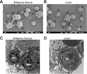
FIG. 5.
The influence of obesity on CD11c+PDCA-1− cells in AT and liver. A and B: SVC from AT and mononuclear cells from liver were isolated and the CD11c+PDCA-1− fraction prepared for SEM. Although macrophage morphology is evident (arrowheads), cells displaying the prominent cytoplasmic veils characteristic of DC are also seen (arrows). C and D: SVC from AT and mononuclear cells from liver were isolated and the CD11c+PDCA-1− fraction prepared for transmission electron microscopy as described (15). Both micrographs show the presence of typical dendritic processes on DC, which are absent on macrophages (Mac), and few vacuoles in DC compared with numerous vacuoles in macrophages.

