FIG. 7.
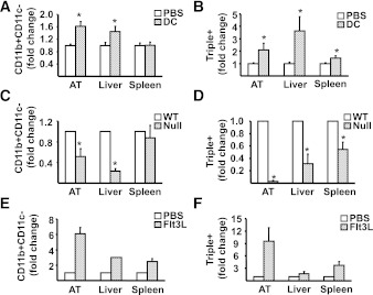
The effects of gain-and-loss of DC on AT and liver macrophage and triple-positive cell infiltration. A and B: 1 week after IP injection of 0.5–1.0 × 10⁶ CD11c+ BMDC in 200 μL PBS (DC) or PBS alone (PBS), mononuclear cells from liver, spleen, and SVC from AT were isolated from mice fed the SCD, stained for CD11b, CD11c, and F4/80 markers, and analyzed by flow cytometry for CD11b+CD11c− and CD11b+CD11c+F4/80+ (triple+). Results are presented as means ± SE (n = minimum of 6 animals/group). Significant differences are indicated (*P < 0.05). C and D: Wild-type (WT) and Flt3l−/− (Null) mice were fed HFD for 16 weeks before isolation of mononuclear cells from liver, spleen, and SVC from AT for analysis by flow cytometry for CD11b+CD11c− and CD11b+CD11c+F4/80+ (triple+). Results are presented as means ± SE (n = minimum of 5 animals/group). Significant differences are indicated (*P < 0.05). E and F: Flt3l−/− mice were injected IP with either 10 μg human recombinant Flt3 ligand in 100 μL PBS (Flt3L) or PBS alone (PBS) every other day for 2 weeks. Subsequently, mononuclear cells from liver, spleen, and SVC from AT were isolated and stained for analysis by flow cytometry for CD11b+CD11c− and CD11b+CD11c+F4/80+ (triple+). Results are presented as means ± SD (n = 2 animals/group).

