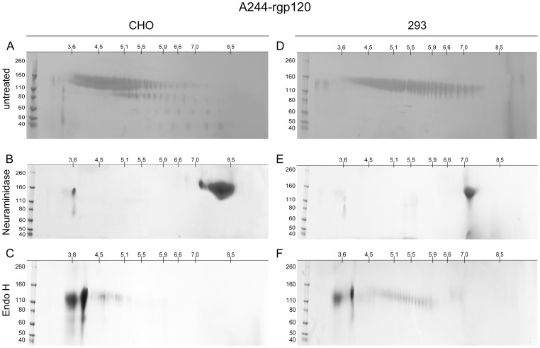
Figure 4. Isoelectric focusing of A244-rgp120 expressed in CHO and 293 cells with and without glycosidase digestion.
The recombinant HIV-1 envelope proteins were left untreated (panels A and D), or digested with neuraminidase (panels B and E) or Endo H (panels C and F). For fractionation in the first dimension, the proteins were resolved by isoelectric focusing using 11 cm IPG strips (pI 3 to 10) (Bio-Rad Laboratories, Hercules, CA). For fractionation in the second dimension, the proteins were resolved using 4–15% tris glycine SDS-PAGE gels. Bands were visualized by Coomassie blue staining. Pre-stained molecular weight markers are shown to the left.

