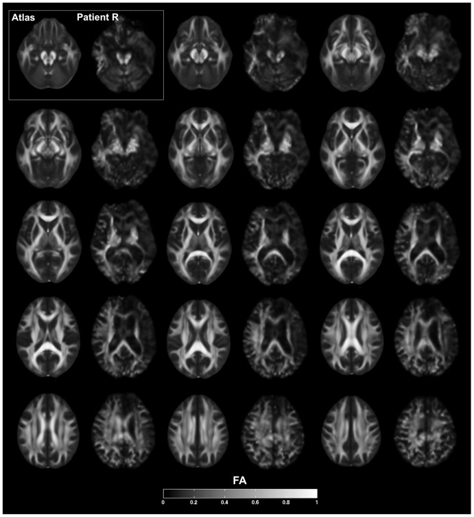
Figure 3. Fractional anisotropy.
Series of axial slices organized in a ventral-to-dorsal direction (ventral-most = top left; dorsal-most = bottom right, as in Figure 2) showing fractional anisotropy (FA) from DTI. Slices are grouped in series of two, corresponding respectively to average values from the FMRIB58 FA atlas and R's brain. Slices are sampled every 4 mm. Slices are in neurological convention, with the left hemisphere on the left. The legend at the bottom of the figure shows the grayscale coding of the degree of FA, ranging from 0 to 1. The results indicate a diffuse and global relative reduction in FA, indicative of extensive white matter pathology and widespread disconnection, especially in the frontal and temporal lobes. There is greater degree of damage in the right hemisphere.

