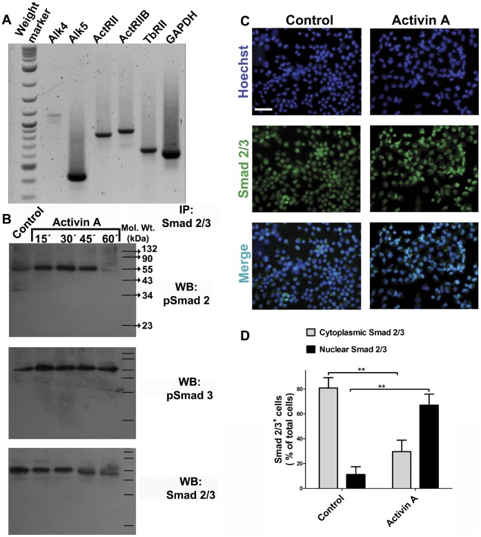
Figure 1. Cerebrocortical neural progenitor cells are responsive to addition of Activin A.
Passage 2 neural progenitor cells (NPC) were obtained from E14 rat embryos and kept in proliferation with FGF2. A: RNA was isolated from these cultures to perform RT-PCR to detect Activin and TGF-β type I and type II receptors. B: Cells were stimulated with 3 ng/ml Activin A at the indicated times. Protein from each sample was obtained and immunoprecipitated with anti-Smad 2/3 antibodies, followed by immunoblot detection of phosphorylated (p)Smad 2, pSmad 3 and total Smad 2/3. The molecular weight (Mol. Wt.) markers are indicated on the right side. C: Immunocytochemistry for Smad 2/3 in cells either untreated or treated during 30 minutes with Activin A. D: Quantification of nuclear translocation of Smad 2/3 from 10 random fields. The values are represented as mean ±S.D. expressed as the percent of total cells (detected by Hoechst staining in blue) with predominant nuclear staining for Smad 2/3 (green label). **p<0.01 versus untreated condition. Scale bar = 50 µm.

