
Figure 2. Nucleocytoplasmic shuttling of VP19C.
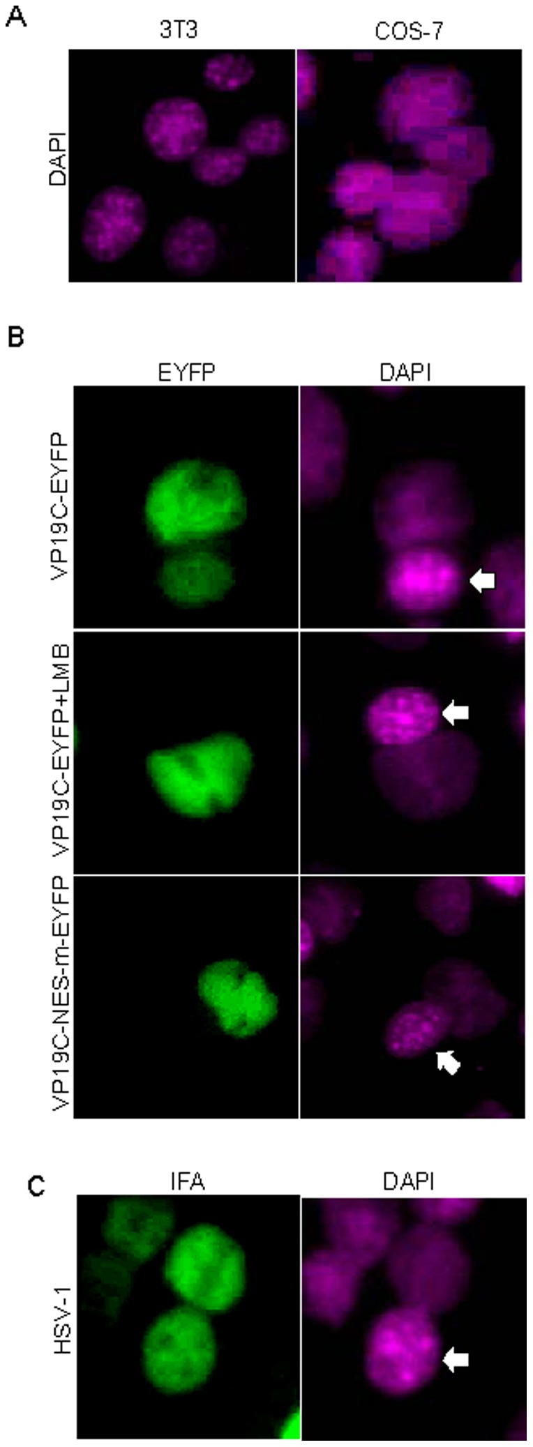
(A) DAPI staining differentiates monkey (COS-7) and murine (3T3) nuclei. (B) Nucleocytoplasmic shuttling of VP19C via a leucine-rich NES: COS-7 cells were transfected with plasmid VP19C-EYFP or VP19C NES-m-EYFP. 24 hours later, transfected cells were subjected to the interspecies heterokaryon assay. Mouse 3T3 cells were identified by their speckled nuclei when stained with DAPI. (C) Nucleocytoplasmic shuttling of VP19C during HSV-1 infection. COS-7 cells were firstly infected with HSV-1 at MOI of 2. At 12 hour after infection, the HSV-1 infected COS-7 cells were mixed with 3T3 cells and subjected to the heterokaryon assay. Mouse 3T3 cells were identified by their speckled nuclei when stained with DAPI. Each image is representative of the vast majority of the cells observed.