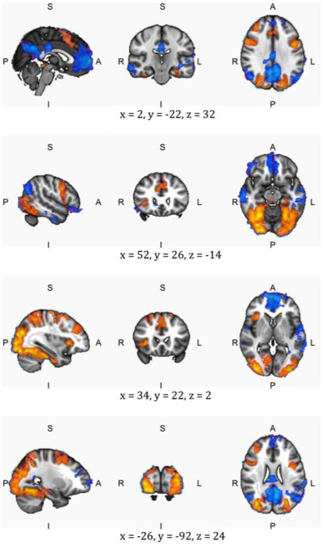
Figure 4. A side, sliced and top-view of four points in the brain: Blue shows the areas that are more active during the familiar condition and red those areas that are more active during the unfamiliar condition.

Center of gravity coordinates (MNI reference system) are shown below each slice. The left picture shows the side view of the brain (s indicates the top of the brain, I the bottom, p the back, and a the front). The middle picture shows a sliced view of the brain (r denotes the rights side of the brain, l the left side). The right picture shows a top view of the brain. For more brain slices: see Appendix S1.
