
TABLE 1. Ligand binding properties of wild-type, E161A, and E169Q human A2a, receptors.
Agonist and antagonist binding affinities (Ki, values) were determined in [3H]CGS 21680 (15 nm) competition binding studies using membrane homogenates prepared from transiently transfected COS-7 cells, as described in Experimental Procedures. Ki, values were calculated from IC50 value with the Kaleidagraph program. Approximately 15 μg of membrane protein/tube were used. The following Kd (nm) and Bmax values for [3H]CGS 21680 (pmol/mg protein, in parentheses) were determined: wild-type, 22.3 ± 4.6 (15.5 ± 0.1); E161A, 41.7 ± 9.2 (17.0 ± 2.7); and E169Q, 57.0 ± 1.8 (9.26 ± 0.37). Values are mean ± standard error of two or three independent experiments, each performed in duplicate.
| ligand | Position of substitution (agonists) |
Ki
|
||
|---|---|---|---|---|
| Wild-type | E161A | E160Q | ||
| nm | ||||
| Agonist 2-CADO 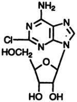
|
2 | 144 ± 33 | 221 ± 71 | 946 ± 20 |
DPMA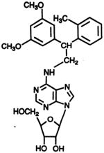
|
N6 | 36.3 ± 1.2 | 61.2 ± 0.2 | 1.62 ± 0.94 |
R-N6-Phenyiisopropyladenosine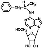
|
N6 | 158 ± 19 | 264 ± 21 | 33.9 ± 2.8 |
| Agonist IB-MECA 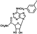
|
N6,5′ | 456 ± 56 | 433 ±13 | 53.4 ± 2.6 |
NECA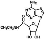
|
5′ | 11.4 ± 4.0 | 23.9 ± 0.2 | 253 ± 40 |
| Antagonist CGS 15943 
|
1.71 ± 0.28 | 0.287 ± 0.16 | 2.26 ± 1.4 | |
| XAC |
6.98 ± 1.6 | 7.19 ± 1.70 | 11.5 ± 2.8 | |