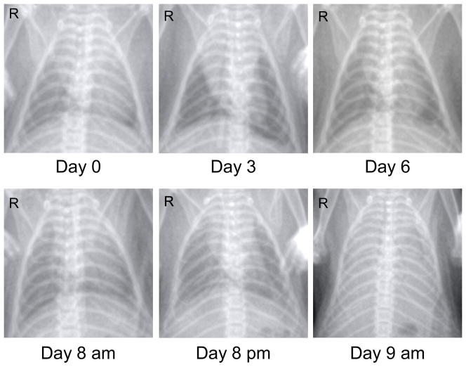
Figure 1. Thoracic imaging of HPS disease progression in a Syrian hamster.
Digital radiography (x-ray) is an important, non-invasive technique which can be utilized to monitor progression of lung infiltrates in Syrian hamsters. Shown are the ventral-dorsal chest radiographs from a single hamster infected with Andes virus (200 focus-forming units, intraperitoneal route). Note the rapidity of lung infiltration which coincides with the onset of severe breathing insufficiences in hamsters (24–36 hours prior to death). The final x-ray taken at the time of euthanasia demonstrates diffuse pulmonary infiltrates with moderate to severe pleural effusion.

