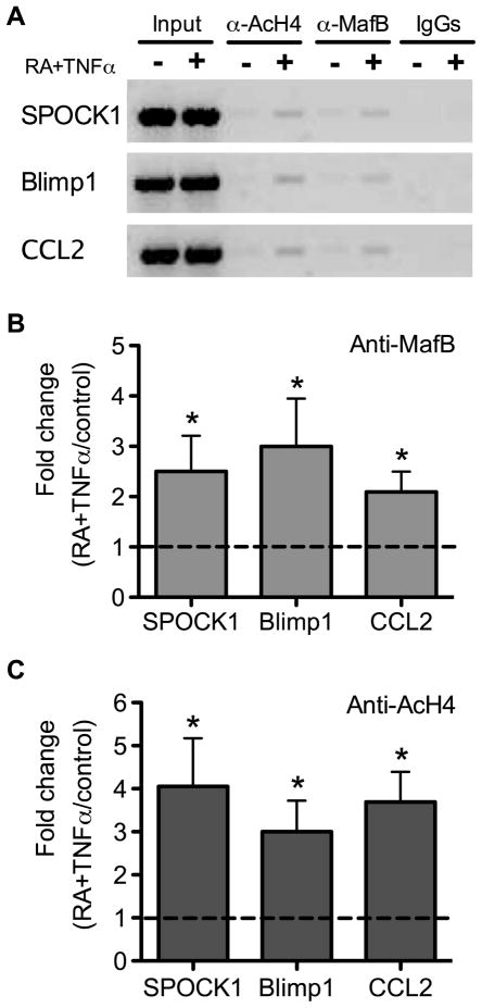
Fig. 5. MafB binding events to the promoter regions of SPOCK1, Blimp1 and CCL2 are enhanced by RA and TNFα.
(A) PCR analysis of DNA after chromatin immunoprecipitation with antibodies to MafB, acetylated histone H4, and IgG as control. Results are shown for SPOCK1, Blimp1, and CLL2 promoter regions after precipitation with anti-MafB antibody (B) and anti-acetylated histone 4 (Anti-AcH4) antibody (C) in ChIP assays. MARE-containing regions were amplified by q-PCR using primers given in Materials and Methods. The Y-axis in B and C represents the fold changes of MafB binding events to MARE-containing regions for cells treated with 20 nM RA plus 5 ng/ml TNFα compared with vehicle-treated control cells (defined as 1.00). Results represented four independent experiments. * P < 0.05 compared to vehicle-treated control cells.

