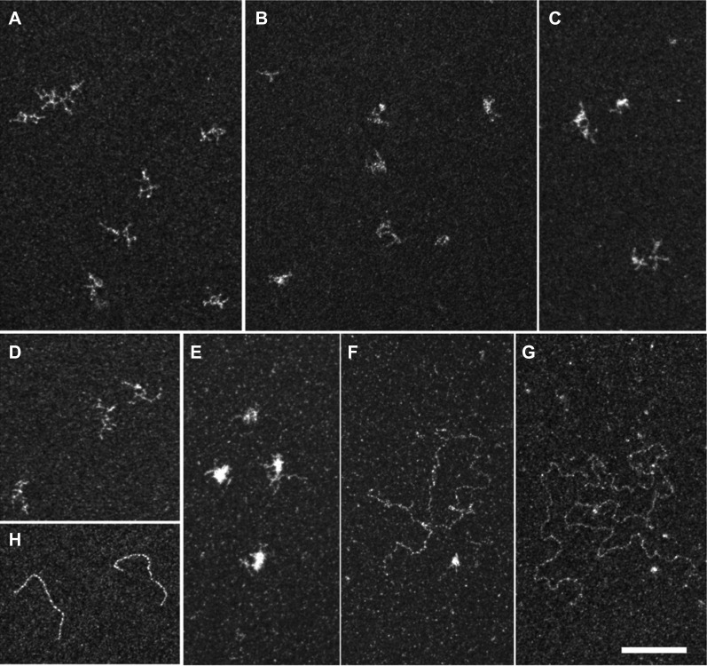
FIGURE 6.
Electron microscopy imaging of RNA bound to Rck/p54. RNA was incubated alone (A,D) or with a 40 molar excess of the double-purified CBP-p54-His (B,C,E–G), in the absence (A–C) or presence (D–G) of ATP. The mixture was spread on grids and stained with uranyl acetate prior to observation by electron microscopy in filtered crystallographic dark field mode. RNA molecules alone were highly branched, independently of the presence of ATP (A,D). Upon protein binding in the absence of ATP, only branched RNA molecules were observed, as seen in two representative fields (B,C). In the presence of ATP, a mixture of ∼80% branched (E) and 20% relaxed (F,G) RNA molecules were observed. The characteristic spreading of double-stranded DNA in the same condition is shown for comparison (H). Magnification ×69,100; bar, 100 nm.

