Figure 2.
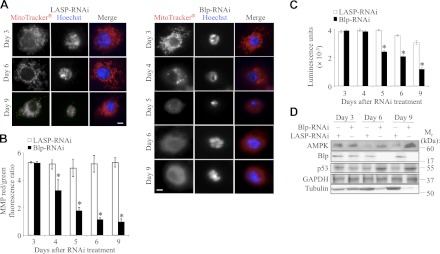
Blp deficiency in S2 cells leads to loss of MMP, reduced cellular ATP levels, and elevation of AMPK. A) Immunofluorescence images of LASP-RNAi and Blp-RNAi-treated cells stained with MitoTracker Red CMXRos to visualize active mitochondria and Hoechst stain for nuclei at different times after RNAi treatment. A heterogenous population was observed, showing slight reduction at d 4 and drastic reduction from d 5 in MitoTracker Red CMXRos staining after Blp-RNAi treatment. B) Ratio of red:green fluorescence after JC-1 staining of RNAi-treated S2 cells, showing loss of MMP from d 4 of Blp-RNAi treatment. C) Cellular ATP levels measured by luciferin-luciferase assay, showing significant reduction of ATP-levels in Blp-RNAi-treated cells from d 5 compared to LASP-RNAi-treated control cells. Each data point is normalized to DAPI staining (DNA). D) Western blot of cell lysates showing a Blp-RNAi-induced increase in AMPK and p53 expression and a decrease in tubulin levels compared with GAPDH (loading control) at d 6 and 9. Scale bars = 20 μm. Values are means ± sd; n = 3. *P < 0.001; Student's t test.

