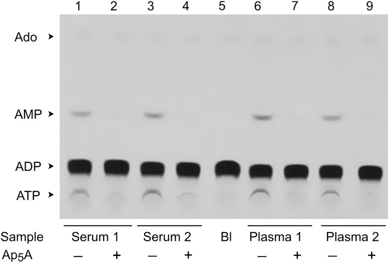
Figure 4.
Autoradiographic analysis of [3H]ADP metabolism in human blood. Serum and heparinized plasma were collected from venous blood of 2 volunteers. Blood samples (10 μl) were pretreated for 20 min in the absence or presence of 100 μM Ap5A prior to addition of 50 μM [3H]ADP. After 60 min incubation, aliquots of the reaction mixture were separated by TLC and developed by autoradiography. Blank (Bl) shows the radiochemical purity of [3H]ADP. Arrows indicate the positions of nucleotide standards and adenosine.

