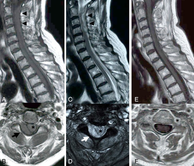
Figure 1.
Cervical MRI in case 1. Sagittal T1- (A) and T2- (C) weighted MRI immediately after the onset showed a longitudinal posterior epidural hematoma from C2 to Th4 (arrow head). Axial T1- (B) and T2- (D) weighted MRI immediately after the onset showed an ovoid epidural hematoma (arrow) in the right postero-lateral aspect and spinal cord (asterisk) compression. T1-weighted sagittal (E) and axial (F) with contrast MRI showed that the hematoma disappeared without abnormal enhancement following conservative treatment at 1 week after the onset.

