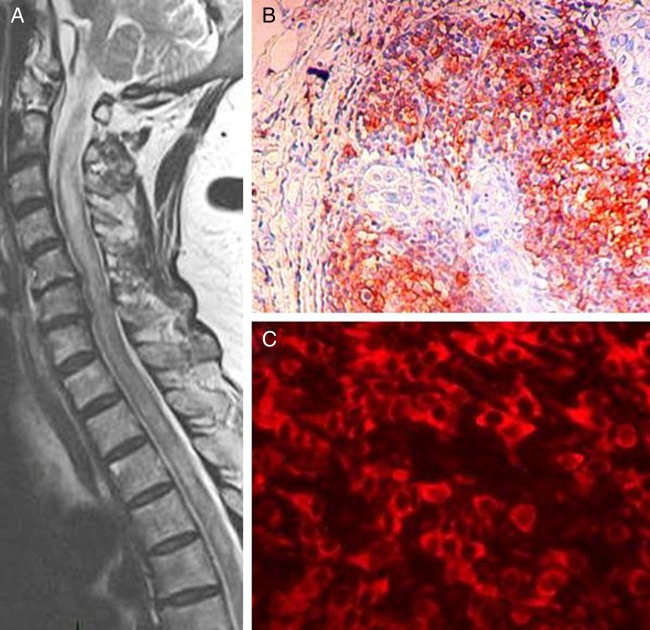
Figure 1.
Sagittal T2-weighted MRI showing long extensive spinal cord lesion at C2–C6 (A) breast tumor section exhibiting CD20+ B cells (avidin–biotin–peroxidase technique with mild hematoxylin counterstaining) (B) aquaporin-4 expressing cells (C, red). B and C panels, original magnification ×100 and ×400, respectively.

