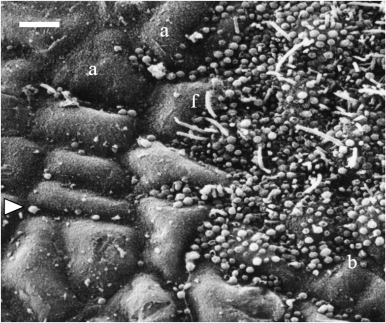
Figure 1.
Scanning electron micrograph showing a partially fouled surface of Fucus vesiculosus with unobstructed and masked areas of host tissue. The left side of the picture shows an apparently clean surface, the algal cells are visible (a) and also few coccoid bacteria (arrow) between them. In contrast, the right side of the picture shows a microbial film with coccoid bacteria (b) and filaments (f) covering the algal cuticle. The photo also illustrates the patchiness of microfouling on one host individual. Scale bar = 5 μm.

