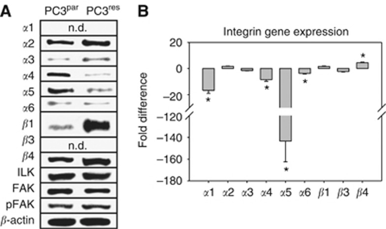
Figure 4.
Modification of intracellular integrin protein level. (A) Lysates of PC3par or PC3res cells were subjected to SDS–PAGE and blotted on the membrane incubated with respective monoclonal antibodies. β-actin served as the internal control. The figure shows one representative from three separate experiments. (B) is related to the integrin gene-expression pattern. Primer sets used for evaluation are listed in materials and methods. Calculation of the relative expression of each gene was done by the ΔΔCt method in the analysis programme of SABioscience Corporation. The housekeeping gene GAPDH was used for normalisation. Values are given as fold difference to PC3par cells. *indicates significant difference.

