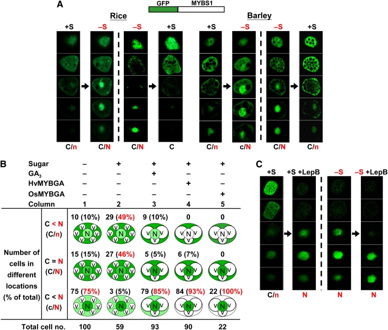
Figure 1.
Glc Inhibits the Nuclear Localization of MYBS1.
Aleurones were transfected with Ubi:GFP-OsMYBS1 and then incubated in +S or −S medium . Thirty optical sections of 0.7 to 0.9 µm thickness each were prepared for each cell; only five regularly spaced sections (sections 3, 9, 15, 21, and 27) are shown here. C and N indicate higher GFP signals and c and n indicate lower GFP signals in the cytoplasm and nucleus, respectively. See also Supplemental Figures 1A and 1D online.
(A) Transfected rice or barley aleurones were incubated in +S or −S medium for 24 h, then shifted from +S to −S medium or from −S to +S medium, and incubated for another 24 h.
(B) Barley aleurones were transfected with Ubi:GFP-OsMYBS1 only or with Ubi:GFP-OsMYBS1 plus Ubi:OsMYBGA and incubated in +S or −S medium. GFP was detected after 24 h. Percentages indicate the number of cells with GFP distribution in the indicated category divided by the total number of cells examined. C, cytoplasm; N, nucleus; V, vacuole. Vacuolation was observed in cells treated with GA3 or overexpressing MYBGA.
(C) Transfected rice aleurones were incubated in +S or −S medium for 24 h. LepB was then added, and aleurones were incubated for another 24 h.
[See online article for color version of this figure.]

