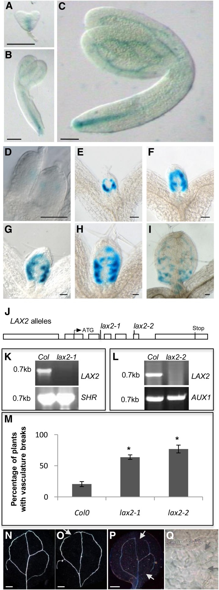
Figure 3.
The lax2 Mutant Exhibits Vascular Patterning Defects in the Cotyledons.
(A) to (C) Promoter:GUS analysis of LAX2 expression in heart stage (A), torpedo (B), and mature (C) embryos.
(D) to (I) Promoter:GUS analysis of expression of LAX2 in developing leaf primordia.
(J) Structure of the LAX2 with the positions of the lax2 mutant alleles indicated. Boxes represent promoter, 5′, and 3′ untranslated regions and exons; lines represent introns.
(K) and (L) RT-PCR analysis of lax2-1 (K) and lax2-2 (L) alleles showing that LAX2 cDNA is detectable in the wild type (Col-0) but not in lax2-1 (K) and lax2-2 (L). Positive controls SHR (K) and AUX1 (L) are detected both in wild-type (Col-0) and lax2 alleles (n = 2).
(M) Graph showing the frequency of vascular breaks in cotyledons of lax2 mutant alleles compared with the wild type (Col-0). Error bars represent se. * indicates statistically significant difference compared with the wild type (Col-0); n = 30; Student’s t test, P < 0.01.
(N) to (P) Differential interference contrast images of wild-type (N), lax2-1 (O), and lax2-2 (P) cotyledons showing the vascular defect in lax2.
(Q) High-magnification differential interference contrast image pinpointing vascular break in a lax2 cotyledon.
Bars in (A) to (C) = 40 μm; bars in (D) to (I) = 100 μm; bars in (N) to (P) = 200 μm.

