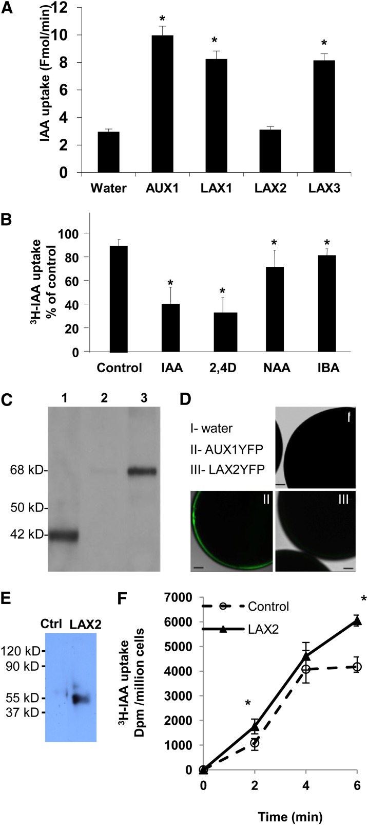
Figure 4.
AUX/LAX Proteins Are Functional Auxin Influx Transporters.
(A) Uptake of [3H]IAA into X. laevis oocytes injected with water or AUX1, LAX1, LAX2, and LAX3 cRNAs at pH 6.4. Oocytes injected with AUX1, LAX1, and LAX3 cRNAs showed increased [3H]IAA uptake when compared with the water-injected control (n = 8).
(B) Uptake of [3H]IAA into oocytes injected with LAX1 cRNA was examined in the presence of excess unlabeled IAA, the auxin analogs 2,4-D and 1-naphthalene acetic acid (NAA), and the naturally occurring auxin form indole butyric acid (IBA) (n = 5).
(C) Immunoblot analysis of oocytes injected with LAX2 (lane 1) or LAX2-YFP (lanes 2 and 3) cRNAs. Total oocyte extract expressing LAX2 (lane 1) or LAX2-YFP (cytosolic fraction, lane 2; microsomal fraction, lane 3) were separated by SDS-PAGE and immunodetected using anti-LAX2 antibodies (dilution 1/1000). Note the size difference between native LAX2 (42 kD) and LAX2-YFP (68 kD).
(D) Laser scanning confocal images of oocytes injected with water (I), AUX1-YFP cRNA (II), or LAX2-YFP cRNA (III).
(E) Immunoblot analysis of empty vector control or LAX2 expressing S. pombe cells. Proteins were separated by SDS-PAGE and immunodetected using anti-LAX2 antibodies (dilution 1/1000).
(F) Uptake of [3H]IAA into empty vector control (dashed line) versus LAX2-expressing (solid line) S. pombe cells compared with zero time point.
Error bars represent sd. * indicates statistically significant difference. Student’s t test P < 0.05.
Bar in (D) = 100 μm.
[See online article for color version of this figure.]

