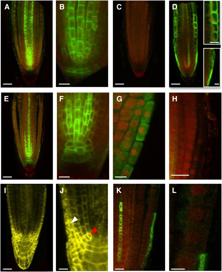
Figure 6.
LAX2 and LAX3 Cannot Be Correctly Targeted in AUX1-Expressing Cells.
(A) to (C) In situ immunodetection of LAX2 (green) in the wild type ([A] to [B]) or lax2 (C) primary roots counter stained with propidium iodide (red).
(D) In situ immunodetection of NHA-AUX1 in root apex. Inset: Close-up of epidermal (Top) and LRC (Bottom) cells.
(E) to (H) In situ immunodetection of LAX2 in aux1-22 ProAUX1:LAX2 roots showing targeting defect of LAX2 in AUX1-expressing cells, including LRC (G) and epidermal cells (H).
(I) and (J) Confocal imaging of seedlings expressing LAX2-YFP under the control of CaMV35S promoter showing correct targeting of LAX2 in LAX2-expressing cells (red arrowhead) but not in AUX1-expressing cells (white arrowhead).
(K) and (L) In situ immunodetection of LAX3-FLAG in aux1-22 ProAUX1>>LAX3 (Methods) roots, showing targeting defects of LAX3 in AUX1 expression domains including LRC and epidermal cells (L).
Bars in (A), (C) to (E), and (I) = 25 μm; bars in (B), (F) to (H), (J), and (K) = 10 μm; bars in (L) and insets = 5 μm.

