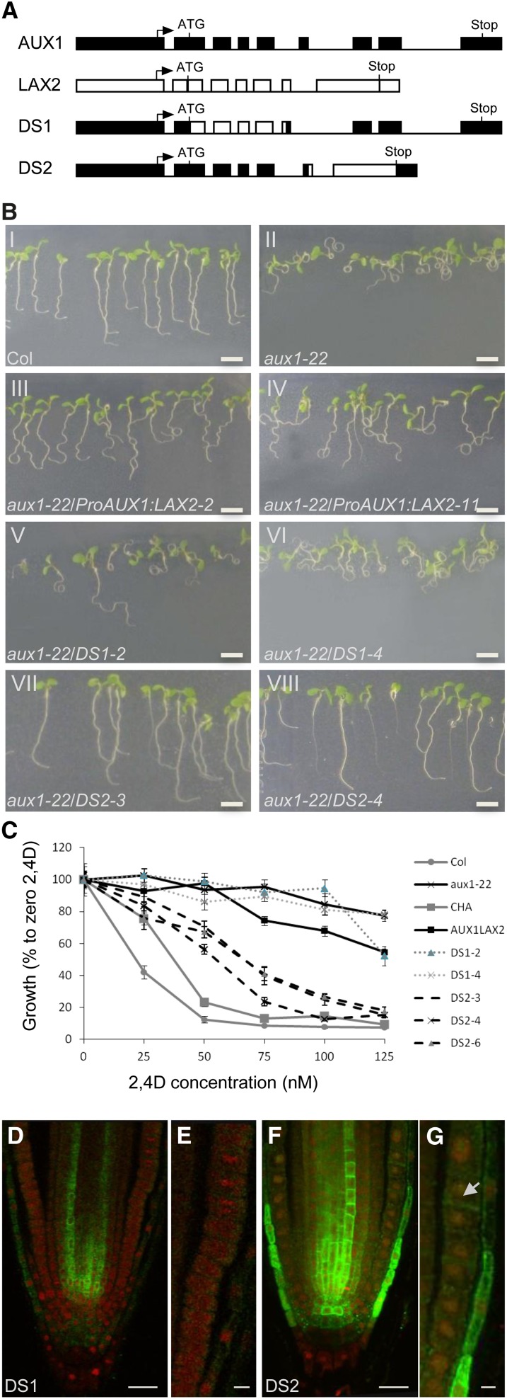
Figure 7.
N-Terminal Half of AUX1 Is Required for Correct Localization in the AUX1 Expression Domain.
(A) Gene constructs used for domain swap experiments (boxes represent promoter, 5′, and 3′ untranslated regions and exons; lines represent introns).
(B) Root gravitropic responses of the wild type (Col-0), aux1-22, and aux1-22 complemented by ProAUX1:LAX2, DS1, or DS2 transgenes (n = 40).
(C) Growth responses of the wild type (Col-0), aux1-22, CHA-AUX1 (CHA), and aux1-22 complemented by ProAUX1:LAX2, DS1, or DS2 transgenes grown at various concentrations of 2,4-D (n = 40). Error bars represent se.
(D) In situ immunodetection of chimeric DS1 protein (green) by anti-HA antibody in primary roots counter stained with propidium iodide (red).
(E) Close-up of LRC and epidermal cells in DS1 roots.
(F) In situ immunodetection of chimeric DS2 protein (green) by anti-LAX2 antibody in primary roots counter stained with propidium iodide (red).
(G) Close up of LRC and epidermal cells in DS2 roots showing localization of DS2 protein (green) in epidermal (arrow) and LRC cells.
(E) to (H) Expression profile of ProLAX1:LAX1-VENUS [(E) to (I)] and ProLAX2:LAX2-VENUS [(J) to (N)] during lateral root primordium development.
Bar in (B) = 5 cm; bars in (D) and (F) = 20 μm; bar in (G) = 5 μm.

