Figure 8.
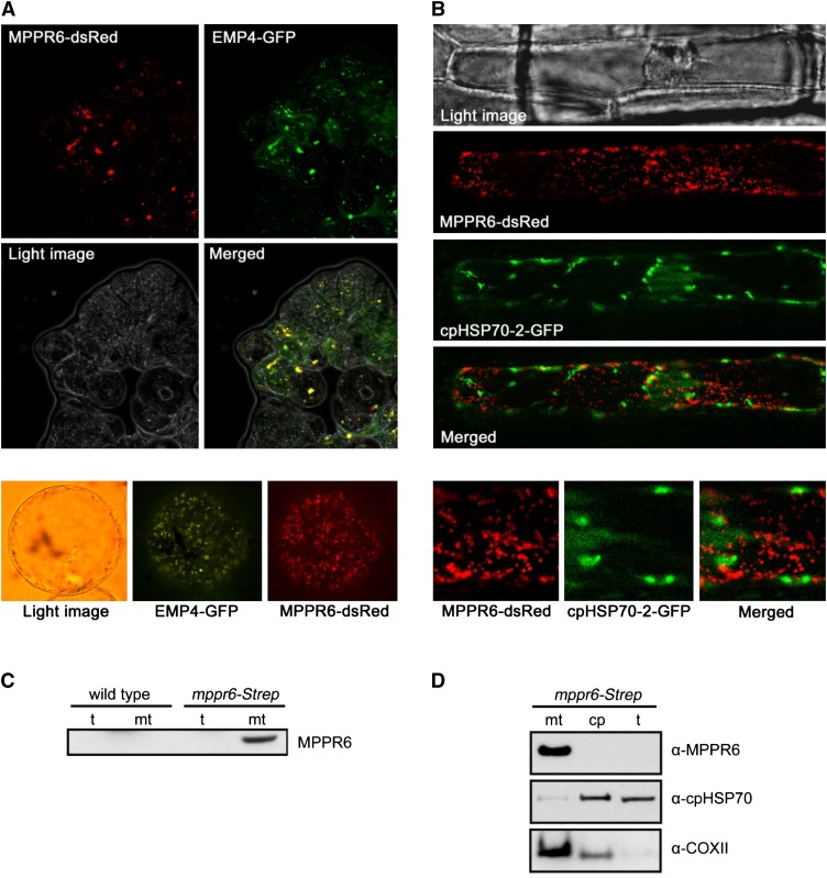
MPPR6 Is Targeted to Mitochondria.
(A) Subcellular localization of MPPR6 in BMS maize suspension cells. Cells were biolistically cotransformed with Ubi:mppr6-dsRed and Ubi:emp4-GFP. Micrographs show red fluorescence signals of MPPR6-dsRed, green fluorescence signals of EMP4-GFP, light image of BMS cells, and merged fluorescence of MPPR6-dsRed and EMP4-GFP. Close-up of a single transformed cell is shown below. Due to the quick movement of mitochondria within the cell caused by cytoplasmic streaming, no precise merging of both signals was possible in the close-up.
(B) Transient coexpression of MPPR6-dsRed and cpHPS70-2-GFP in onion epidermis cells. A merged image of the nonoverlapping fluorescence is shown. Close-ups of selected regions are shown below.
(C) Immunodetection of the MPPR6 protein in total protein (t) and crude mitochondria extract (mt) obtained from wild-type and mppr6-Strep overexpressing maize seedlings.
(D) Immunoblot analyses detected the MPPR6 protein in mitochondria but not in chloroplast (cp) fractions, both obtained from the mppr6-Strep overexpressor. The same filter was reprobed with chloroplast- (α-cpHPS70-2) and mitochondria-specific (α-COXII) antibodies.

