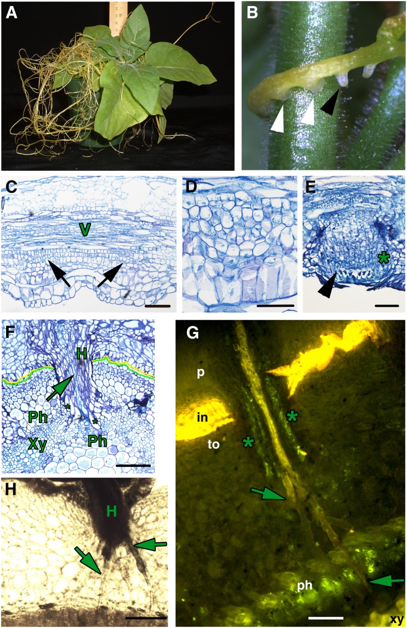
Figure 1.
Parasitism by Dodder.
(A) Tobacco infected with dodder.
(B) Dodder strand showing embedded haustoria (white arrowheads) and prehaustoria (black arrowhead).
(C) Longitudinal section of a dodder strand with vascular tissue (V) and initiating haustoria (black arrows).
(D) Longitudinal section of a dodder strand in high magnification showing developing haustoria.
(E) Dodder haustoria emerging from the stem epidermis; the asterisk marks file cells, and the arrowhead marks digitate cells in the prehaustorium.
(F) Transverse section of a tobacco stem with an attached dodder strand. Mature haustoria (H) of dodder (marked with a yellow line) are embedded in the tobacco stem (demarcated with a green line) and Xylic searching hyphae (arrow) have contacted the host xylem (Xy), while Phloic hyphae (asterisks) contact the host phloem (Ph).
(G) Transverse section of a tobacco stem (to) with attached dodder parasite (p) stained with Aniline Blue. Mature haustorium shows a central column of Xylic hyphae (arrows) that have contacted the host xylem, and peripheral to that, Phloic hyphae (marked with asterisks) that contact the host phloem. in, host-parasite interface.
(H) Transverse section of a tobacco stem with attached dodder parasite showing a haustorium sending out numerous searching hyphae (arrows) toward the host vascular tissue.
Bars = 100 µm.

