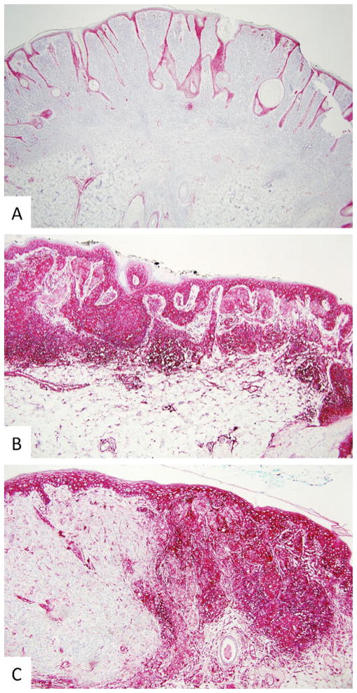
Fig. 4.
Differential HLA antigen expression in benign and atypical melanocytic nevi, in cutaneous melanoma and in surrounding normal cells. a Only normal keratinocytes, endothelial cells, and antigen-presenting cells were marked by HLA class I heavy chain-specific mAb HC-10 in the immunoperoxidase reaction in an intradermal nevus (×10). b Normal keratinocytes, endothelial cells, antigen-presenting cells, and melanocytes were marked by HLA class I heavy chain-specific mAb HC-10 in the immunoperoxidase reaction in a severely atypical nevus (×10). c Normal keratinocytes, endothelial cells, and malignant melanocytes but not intradermal nested melanocytes or vertical growth phase melanoma cells were marked by HLA class I heavy chain-specific mAb HC-10 in the immunoperoxidase reaction in a superficial spreading melanoma arising within an intradermal nevus (×10)

