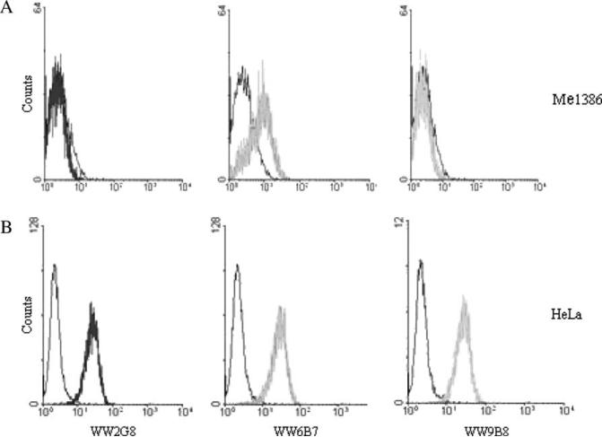
Figure 5.
Characterization of major histocompatibility complex class I chain-related gene A (MICA) monoclonal antibodies (mAbs) against MICA-positive and -negative cells by flow cytometry. HeLa or Me1386 cells (5 × 10⁵) were incubated with anti-MICA mAbs and followed by fluorescein isothiocyanate-labeled rabbit anti-mouse F(ab′)2. Stained cells were analyzed in FACScan equipped with Cell-Quest™ software. Analysis of WW2G8, WW6B7 and WW9B8 against Me1386 (A) and HeLa (B) cells by flow cytometry. Isotype controls are shown in black.

