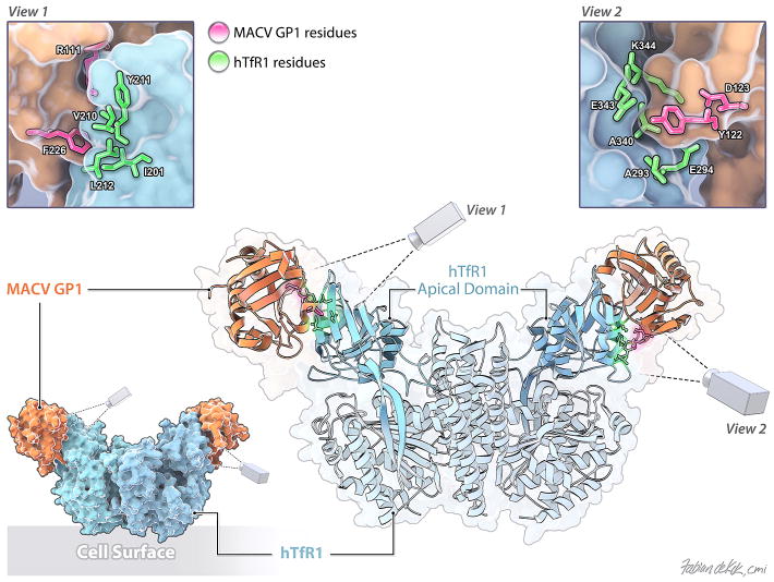
Figure 3.
Structure of the MACV GP1-hTfR1 complex (as described in192) with the cell surface orientated to the bottom (PDB ID number: 3KAS). The TfR1 apical domain is colored cyan. MACV GP1 is colored in apricot. Two enlarged views of the TfR1:MACV GP1 contact sites are shown. MACV residues important for TfR1 binding are labeled and colored in magenta. TfR1 residues are labeled and colored in bright green.

